By Mike Pablo
Today I’m writing about microscopy. One of the great pleasures of the field of microscopy is being able to look at cool pictures, so let’s do that first:
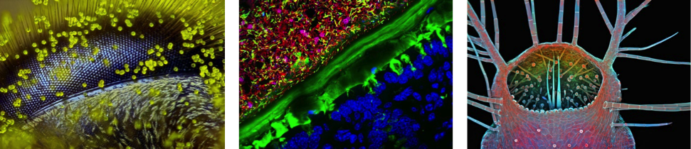
From left to right. The first, second, and third place winners of the 2015 Nikon Photomicrography Competition. Try guessing what they are, then look them up! The first one does something called a “waggle dance”…
Microscopes are among the most iconic symbols of science, alongside other famous images like DNA, test tubes, and the atom. Though magnification devices had existed for many years prior (try reading about the invention of eye glasses in 13th-century Italy and the invention of the first microscopes by 16th-century father-son duo Hans and Zacharias Jansen!), Anton van Leeuwenhoek (1632-1723) is credited as the first to make and use a proper microscope for scientific use. However, his invention looked nothing like the microscopes we use today.
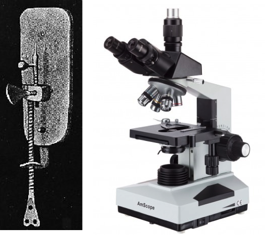
Left: The design of Anton van Leeuwenhoek’s microscope. Right: A modern microscope.
From the University of California Museum of Paleontology and AmScope
Van Leeuwenhoek made groundbreaking discoveries, including the first identification of bacteria and protozoa. He changed the world by seeing what was once unseeable. Building from his work, researchers working on optics and microscope design have allowed scientists to take progressively closer looks at the fine details of the world around us. As you might guess from how completely different van Leeuwenhoek’s microscope looks from a modern microscope, microscopy has changed quite a bit. The definition of what is “unseeable” has changed repeatedly over the years, which has facilitated many new discoveries — but I’m getting a little ahead of myself.
In 1873, about 150 years after van Leeuwenhoek built his microscope, the physicist Ernst Abbe showed that there was a wall in microscopy: there was a fundamental limit to what could be seen. This limit was not because of engineering flaws or our inability to build microscopes, but rather due to the nature of light itself. (It doesn’t apply to other kinds of microscopes, such as electron and atomic force, which I won’t get into here.) Tip-toeing around the physics, the best attainable resolution of a conventional light microscope is about ~250 nm. This has to do with both refraction and the wave nature of light. That is, assuming you could get your hands on extremely high quality lenses, perfectly calibrate it, and so on, light itself, prevents you from distinguishing two objects closer than roughly 250 x 10-9 meters (~0.0000098 inches).
If you were reading carefully, you’ll notice that I specified a conventional light microscope. The recent 2014 Nobel Prize in Chemistry was awarded to three scientists (Betzig, Hell, and Moerner) for “having bypassed a presumed scientific limitation stipulating that an optical microscope can never yield a resolution better than 0.2 micrometres.” (The Nobel Prize in Chemistry 2014 – Popular Information). In other words, the award was for super-resolution microscopy, or ways for optical microscopes to “see” with resolution better than Abbe’s ~250 nm limit, which had stood strong for about a century.
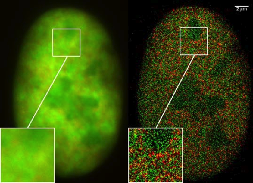
Comparison between conventional microscopy (left) and super-resolution images (right). Objects separated by distances less than 0.2 μm (= 200 nm) are distinguishable in the enlarged image on the right. This is the nucleus of a bone cancer cell. Image from Wikimedia Commons
Before these advances, optical microscopes were important among biologists to ask important questions (what do these cells look like, how many are there, what are they doing…?). Thanks to advances in microscopy, we’re able to answer questions which were once inconceivable to tackle. Since these microscopy techniques allow us to now look at individual molecules in cells, we now can “see” how biochemistry happens at amazing detail.
Big discoveries are waiting to be made down at those little sizes.
If you thought this topic was interesting, or if you want a more technical perspective of the topic, I suggest you take a look (ha!) at the blog post series written by Everyday Scientist, as well as the Nobel prize page Popular and Advanced Information!)
Edited by Anna Chiarella and Tamara Vital