By Anna Wheless
HOW FAR WE’VE COME
“What was the most important scientific discovery ever made?”
The answers to this question range from the discovery of antibiotics to the development of the theory of general relativity. It’s all a matter of opinion, but my answer to this question would be: the manipulation of glass. It sounds so simple, but the ability to precisely make lenses out of glass not only enabled the visually impaired (like myself) to see with glasses, it also enabled the invention of the microscope, which completely changed the way we investigate our world. The professor and author Mark Miodownik has said that glass allowed us to “transcend our scale” and see into the world of microorganisms, and this ability laid the foundation for modern medicine. Without the microscope, we would never have known that our own cells, bacteria, or viruses even exist.
People have been using microscopes since the 1600s to make observations about the world around them. When they were first invented, they looked like the picture you see below. Only the most skillfully made microscopes from this era could be used to view things like animal, plant, and bacterial cells. Today, it is possible to use ultra-powerful microscopes along with advanced software to produce images of proteins, viruses, and small parts of cells.
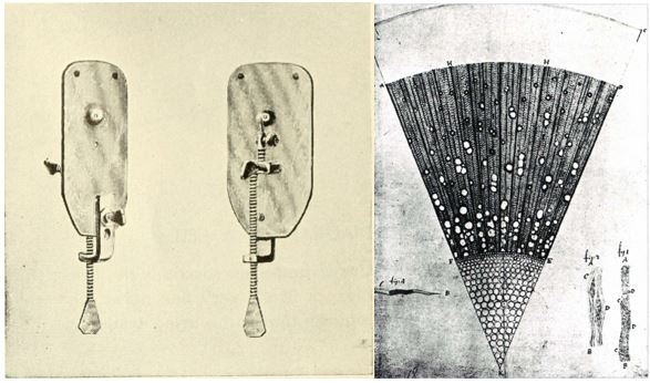
Left: A single-lens microscope designed by Antonie van Leeuwenhoek in the 1600s, about 4-5 inches tall. The greatest magnification possible for these devices is debated, but some estimate it was about 275x. Right: A drawing by Antonie van Leeuwenhoek of a microscopic section of wood.
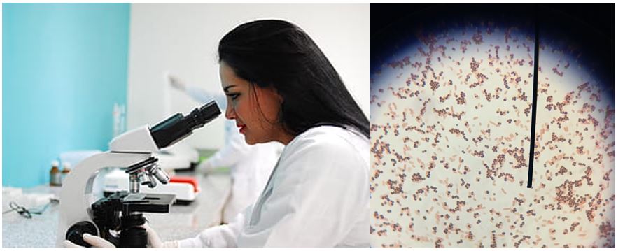
Left: A typical light microscope you might have seen in a lab. Right: Image of bacteria (E. coli and S. epidermidis with Gram stain) taken with a light microscope, total magnification 1000x. (Image is my own)
The microscopes you may have used in your science labs are a type of simple light microscope. They use lenses made out of glass to bend light and magnify the image of your sample, which is similar to how the first microscopes worked. They’re great for looking at tissues and cells if you don’t need to see smaller structures. Fancier types of light microscopes can offer greater detail of cells than your classroom microscope, but with traditional light microscopy you can’t see anything smaller than the wavelength of the light you’re using to see the sample*. Viruses and proteins are generally smaller than this wavelength, so you need another technique to image them.
ENTER: THE ELECTRON MICROSCOPE

Left: An advanced electron microscope, called a Krios, with its side panels open. Right: An image (called a micrograph) of proteins (seen here as tiny rings) taken with electron microscopy.
The super-powerful microscopes that can make detailed images of proteins and viruses are fundamentally different from light microscopes–they don’t use light at all. Instead, they use beams of electrons to detect the boundaries of the molecules you’re imaging. Electrons, like photons, are particles that also have wave behavior, but the wavelength of electrons is much smaller than the wavelength of visible light. The smaller wavelength of the electrons gives these microscopes greater resolution. Inside the microscope, a source of electrons is aimed at the sample. This beam of electrons is focused with lenses, but unlike the lenses in light microscopes, these lenses aren’t made of glass. Instead, they’re wires with electricity running through them–or in other words, electromagnets. The lenses have to bend the path of electrons just like glass lenses bend the path of light, which is exactly what the magnetic field of these lenses will do. A part of the microscope called the detector will transmit data about the electrons to a computer. Finally, a scientist will use this data to try to improve the resolution as much as possible and construct a model of the sample. The result is a picture of something that is only a few billionths of a meter across.
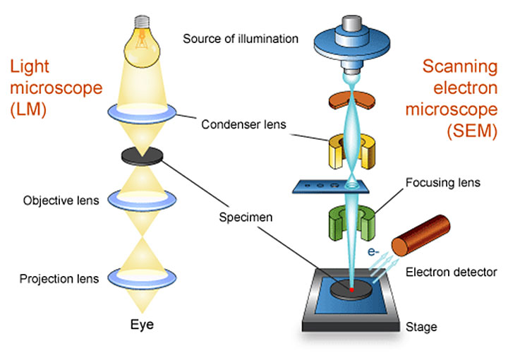
Above: Comparison of a light microscope and a type of electron microscope called a scanning electron microscope. This is a little different from the electron microscope used to image proteins, which is called a transmission electron microscope, but the basic ideas are similar.
The images of tiny structures that we get from electron microscopes can help us understand diseases and design new medications and materials. The first microscopes were used to view cells 350 years ago, and now we can see structures that are much smaller than cells. Who knows what we’ll be able to see in the next 350 years?
*Footnote: recent advances in light microscopy using “super-resolution” techniques have provided another way around the diffraction limit, but still don’t approach the level of structural detail we can achieve with electron microscopy
Edited by Meryem Ok