By Alec Chaves
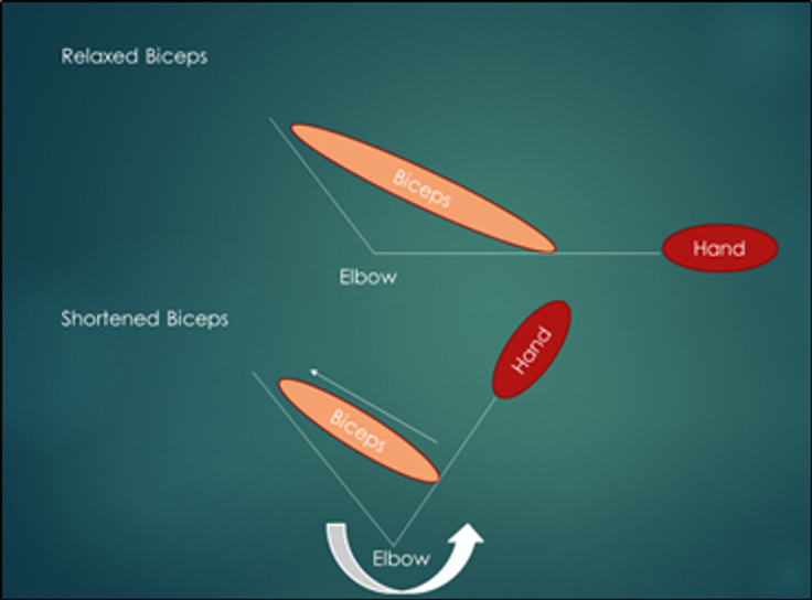 There are many aspects of human biology that are fascinating…. how we breathe, how we turn food into energy, and even the process of forming thoughts. In this article, I am going to break down one of the most basic functions that humans can carry out: movement. Although basic, movement requires a highly coordinated effort from the nervous, skeletal muscle, and skeletal system. To get started, let’s first talk about what the muscle needs to do in order to create movement, and that is to “shorten.” It might seem counterintuitive, so let’s look at our biceps muscle for example. The biceps muscle is found on the front of our arm and attaches to our shoulder and to our forearm. When this muscle “shortens,” it will pull on the forearm moving it closer to your shoulder which ends up looking like a bicep curl (depicted in this diagram).
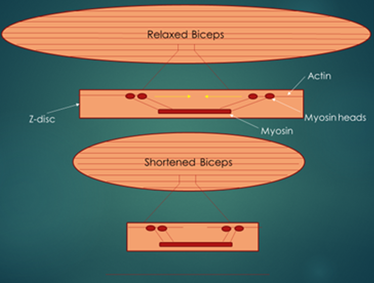
Now, the part about this process that I find most fascinating is what happens inside the muscle so that it can shorten. Like other tissues in our bodies, our muscle is made up of individual cells, which are aligned in series and contain several proteins (i.e. organic structures that provide many functions for us to survive), some of which are designated to help the muscle shorten. Two of these proteins are known as actin and myosin. Actin is a structural protein that is anchored to the ends of a muscle cell known as a z-disc. Myosin, on the other hand, is found in the middle of the muscle cell and has appendage-like structures known as myosin heads. When energy is readily available and our nervous system sends a signal to our muscle, the myosin heads will pull on actin, to move it closer to the middle of the cell. An interesting side note: one of the reasons people with bigger muscles are usually stronger, is because they have a greater amount of myosin heads in their muscle, and therefore have more of them to pull on actin to generate force. As I mentioned before, actin is attached to the z-disc on either side, so when actin moves, it will pull the z-disc along with it. As a result, the two z-discs will move closer to each other, making the muscle appear shorter. Now, imagine this process happening simultaneously in thousands of other cells in ou r muscle. Since these cells are connected in series, each cell will pull on the one next to it so that they all move closer to the middle of the muscle, making the muscle appear shortened/contracted (depicted in second diagram).
 Something I did not mention is that on the ends of muscle, there are connective tissue segments known as tendons. One of the more famous tendons is the Achilles tendon, which connects our calf muscles to our heel. When our muscle shortens, it will pull on the tendon and the bone it is attached to in the same direction that the muscle is shortening in. So, when our calf muscles shorten and pull our Achilles tendon, this will pull the heel upward, allowing you to get on your tippy toes.
Although you cannot see actin and myosin with the naked eye, appreciating their roles in controlling how our bodies move is quite fascinating. If the muscle anatomy and the process of human movement interests you, try using the “shortening” concept to figure out what other muscles in our body are responsible for. For example, try straightening your knee and feel what muscles contract and shorten.
Happy Muscling!
Edited by Keean Braceros and Sarah Brotman